Tag Archives: Storz

Paediatric Bonfils Intubation

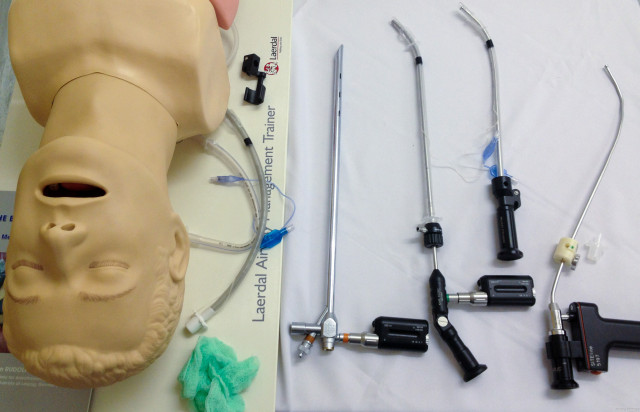
Intubation in a young child with a severe submandibular abscess using the paediatric Bonfils rigid intubating endoscope under inhalational general anaesthesia. Direct laryngoscopy showed only severe swelling with a Cormack-Lehane grade 3b view.  A standard laryngoscope was used with the left hand to create an open path for the Bonfils just right of the midline, avoiding the worst of the submandibular swelling.  3.5mm Bonfils allowed intubation with a 4.5 mm uncuffed ETT.  Note that because this is a rigid intubating scope, it is not inserted through the vocal cords, but they are visible through the tube as it is inserted with the Bonfils held steady.

AirwayHowTo: Leak test a flexible endoscope

Flexible fibreoptic and video endoscopes are fantastic but expensive pieces of equipment. In order to be maintained in top working condition, they need a little tender loving care. A particular problem occurs when the sheath of the scope becomes damaged or cracked, allowing fluid (especially corrosive cleaning solutions) to enter the inner workings of the scope, causing irreparable damage. The inner workings of the scope are a sealed environment. The patency of the seal – and thus the presence or absence of any damage – can easily be determined by performing a leak test. Although this is usually performed by medical technologists who are looking after the scopes, it can just as be performed quickly by the user while the scope is being prepared, or just before cleaning. Spend 100 seconds watching this brief, unadorned video which will walk you through the process. The demonstration here is using our Storz equipment, but is very similar regardless of the make or model of endoscope.

Cormack-Lehane Grading Examples

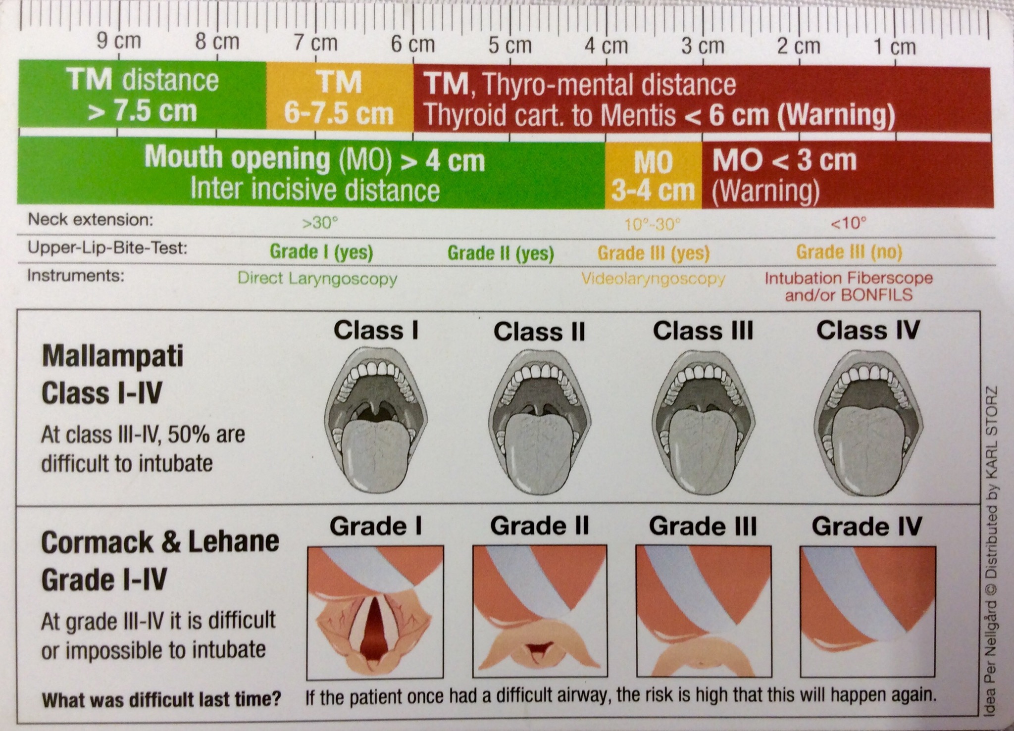
Here are some real-life examples of Cormack-Lehane classification of laryngoscopic view taken with a video laryngoscope.  Although initially described for direct laryngoscopy in obstetric patients, it is a useful descriptive system in many settings, but is frequently misreported and/or misunderstood.  We will continue to expand the set as we collect good images.

Original and revised (Yentis & Lee, 1998) CL grading:

Click to access the paper in Anaesthesiology, 1998

Colour-coded to make it a little easier to read:

…and here from the CoPilot VL material, a more anatomically-correct sketch which shows the distinction between 2a and 2b clearly:

Cormack–Lehane 1:Cormack-Lehane 1

Cormack-Lehane 2a:Cormack-Lehane 2a

Cormack-Lehane 2b:2014.01.16_08.04.59 Cormack-Lehane 2b (nearly 3)

Cormack-Lehane 3:2014.02.03_10.21.59 Cormack-Lehane 3a

Cormack-Lehane 4:2013.09.20_11.19.39_tongue CL4

See the original article by RS Cormack and J Lehane in Anaesthesia, 1994;(39):1105-1111.

Various modifications to this scheme have been proposed for use with video laryngoscopy (VL), including suggestions by Cook and Fremantle:

cry_redusaabnj

freemantly-vl-grading


Bougie-assisted intubation snagging on arytenoid

This common problem is worst with small bougies, such as in this paediatric example of a child with severe burns and a difficult airway.  The bevel of the ETT allows the tip of the tube to stick out right (laterally) of the bougie and snag on the right arytenoid cartilage.  This can be remedied by withdrawing the ETT slightly (to disengage it from the arytenoid), effecting a one-quarter counter-clockwise rotation of the ETT on the bougie (bringing the bevel and tip of the ETT into a superior midline position snug with the bougie), and then advancing again.