Tag Archives: video laryngoscopy

VLVids: Dual endoscopy intubation with compressive thyroid mass

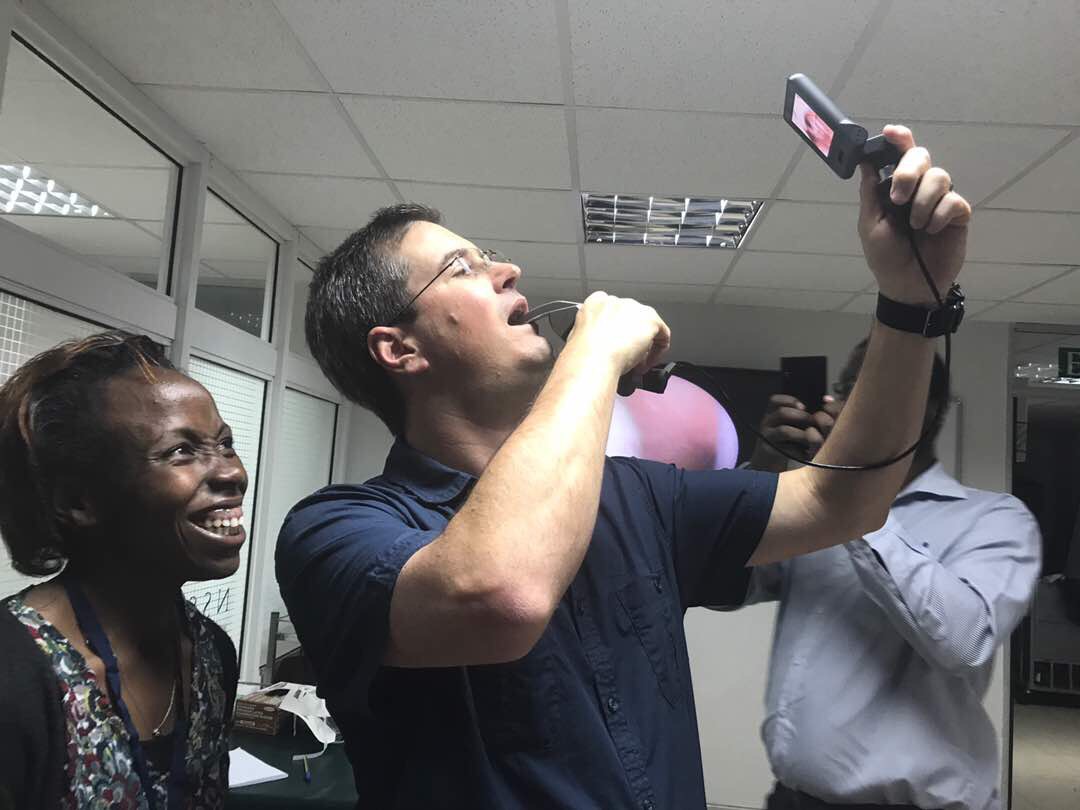
Here’s a great example of using a dual endoscopy technique not only to manage a potentially difficult airway, but also to give more insight into the pathology itself. This short video shows the intubation of a patient who presented for thyroid surgery with a large mass compressing the trachea, as well as some other predictors of difficult intubation: slightly limited mouth opening, a short neck, and potentially challenging dentition. We wanted to see the position and degree of tracheal compression present before advancing the tracheal tube, but also to place the modified tube with nerve monitoring sensor precisely at the right depth.

Video laryngoscopy with a Mackintosh-style blade provided good access to the airway, and allowed us to spray the vocal cords and trachea with local anaesthetic, avoiding neuromuscular blockers. We could then place the tip of the tube through the vocal cords and observe the trachea and subglottic space using the Bonfils optical stylet. Both images are displayed side-by-side to coordinate view. It’s a good tip to put the device in your left hand on the left side of the screen (and corresponding right-handed device on the right) to promote good orientation.

If you’ve tired dual endoscopy for a clinical case, pop a description or brief story in the comments!

Leave a Reply

DAS 2021 – Awake Video Laryngoscopy

Selected references can be accessed online via the links here. If you are unable to access a reference, or wish to download a larger set for furhter reading, please click the link for the archive at the bottom.

If you’re struggling access the articles above or would like an EndNote library and further references, you can access a share folder here.

A brief video describing a challenging case managed with awake VL using the CMAC Video Stylet is shown below. (Video prepared for the 2021 Virtual Guy’s Airway Management Course)

COVID Airways Podcast on Critical Care Scenarios

I was honoured to be invited back on the Critical Care Scenarios (@icuscenarios) podcast, to chat this time about airway management in COVID-19, but also talking about how it’s helping to focus improving our emergency airway management in general. Check it out at this link:

http://icuscenarios.com/episode-37-airway-management-for-covid-19/

Dual endoscopy demo

Here’s a brief clinical example of dual endoscopy using the CMAC VL and VS (video stylet).  This case was performed for teaching purposes, rather than a difficult airway.  Note the endoscopic view appearing as a picture-in-picture is from the editing; the images are on two separate displays.  (It starts a little late due to the author forgetting to press the record button on the device).


Nairobi Airway Course 2017: Photos

Some photos of the action at the Nairobi Surgical Skills Centre this week. Many thanks to the local faculty from the University of Nairobi and other institutions, the companies that supported use of the facilities and equipment, and the enthusiastic delegates!


Resources and notes from the course are available on the course page here

VLVids: Room for Improvement!

This real-life videolaryngoscopy example (RSI of a patient with a head injury for craniectomy) shows some commom errors, and room for improvement. Three major areas that could be better to spot here:

  1. Less-than-ideal positioning of the VL blade. The tip is too deep (over the epiglottis, rather than in the vallecula), which reduces the available space to manipulate both VL and ETT, the field of vision, and the ability to perceive the approach of the ETT.
  2. Quick suctioning of the secretions/saliva/mucus would reduce the risk of losing the view or the patient aspirating.
  3. An introducer (stylet/bougie) would greatly assist passage of the ETT through the cords.

Is fibreoptic intubation a dying art?

Over the past two weeks, I have been involved in three cases where all means of laryngoscopic intubation failed – including multiple different blades, introducers and highly skilled hands – and the airway could only be intubated with a flexible fibreoptic ‘scope.  These three cases illustrate the type of pathology that can make even video laryngoscopy (VL) difficult or impossible:

  • A morbidly obese patient in traction with a high spinal injury
  • A patient presenting with late-stage, advanced laryngeal carcinoma with both supra- and infraglottic involvement and masses
  • A child with Pierre-Robin Sequence presenting for mandibular distraction surgery.

In an article on the Airway E-Learning site, Dr Matthew Wiles details why he thinks fibreoptic intubation (FOI) is becoming a rare beast, and why we should work hard to maintain excellence in this important skill.

Despite being a huge fan and daily user of VLs, I am completely in agreement with his sentiments.

Bougie-assisted intubation snagging on arytenoid

This common problem is worst with small bougies, such as in this paediatric example of a child with severe burns and a difficult airway.  The bevel of the ETT allows the tip of the tube to stick out right (laterally) of the bougie and snag on the right arytenoid cartilage.  This can be remedied by withdrawing the ETT slightly (to disengage it from the arytenoid), effecting a one-quarter counter-clockwise rotation of the ETT on the bougie (bringing the bevel and tip of the ETT into a superior midline position snug with the bougie), and then advancing again.

https://www.youtube.com/watch?v=DvnQL56FHZ8&feature=youtu.be